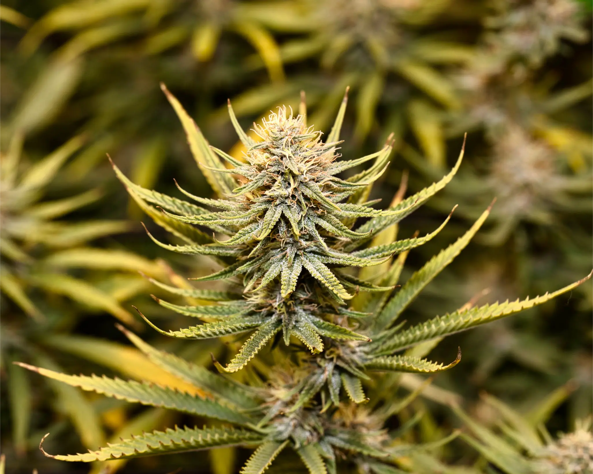
Why Cannabis Leaves Turn Yellow: Diagnosis, Physiology, and Systematic Recovery
Yellowing cannabis leaves (chlorosis) represent the single most common visual alarm in commercial and craft cultivation. However, yellowing is not a singular diagnosis; it is the universal downstream manifestation of chlorophyll degradation. When chlorophyll pigment catabolism outpaces biosynthesis, leaf mesophyll tissue loses its vibrant green color, unmasking yellow carotenoids and xanthophylls. Inexperienced growers frequently assume every yellow leaf signals nitrogen deficiency, reflexively increasing fertilizer strength. This knee-jerk reaction frequently triggers devastating root-zone crises, transforming minor environmental hiccups into severe salt toxicity and systemic nutrient lockout. Yellowing can be triggered by mobile mineral reallocation (nitrogen, magnesium), immobile micronutrient blockades (iron, manganese, sulfur), root-zone hypoxia from overwatering, cold media temperatures (<18 °C), extreme vapor pressure deficit (VPD) drift, light bleaching, or the healthy programmed senescence of late-flowering ripening. This clinical guide breaks down the biochemistry of chlorophyll catabolism, establishes an infallible anatomical diagnostic sequence, models substrate pore-water interactions, and provides a systematic, single-variable protocol to restore canopy vitality.
Direct Answer and Scope: Deconstructing the Yellow Leaf Alarm
When a cannabis leaf turns yellow, the plant is signaling that its rate of chlorophyll degradation has exceeded its rate of chlorophyll biosynthesis. Chlorophyll pigments are unstable organometallic complexes that undergo continuous enzymatic turnover within chloroplast thylakoid membranes. Under steady-state metabolic conditions, the plant continuously synthesizes new chlorophyll molecules to replace those damaged by photochemical light absorption.
Yellowing occurs under two primary biological scenarios:
- Active Programmed Re-Allocation (Source-Sink Translocation): When essential mobile mineral building blocks (particularly nitrogen, potassium, and magnesium) become scarce in root pore water, the plant initiates a survival strategy. It synthesizes catabolic enzymes in older, shaded lower fan leaves, dismantling chlorophyll and chloroplast proteins. The liberated nutrients are loaded into the phloem stream and translocated upward to sustain active cell division in the apical meristems and expanding floral buds. In this context, lower-leaf yellowing is an active, orderly evacuation of resources.
- Stress-Induced Cellular Breakdown (Pathological Chlorosis): When external environmental or chemical stressors – such as substrate waterlogging (hypoxia), root-zone pH lockouts, extreme salinity, or pathogenic fungal attack (Pythium, Fusarium) – disrupt root respiration and transpiration, cell metabolism collapses. Cells can no longer maintain membrane electrochemical gradients, and chlorophyll degrades uncontrollably without nutrient recovery.
By the end of this guide, cultivators will possess the clinical framework to pinpoint the exact root cause of foliar yellowing within three minutes, differentiate true starvation from false-positive environmental stress, and execute a disciplined correction without stacking fertilizers.
Foliar yellowing reflects chlorophyll breakdown, triggered by mobile nutrient remobilization, root hypoxia, pH lockouts, or natural late-flower senescence.
Core Mechanisms: Chlorophyll Catabolism and the PAO Pathway
To accurately read foliar distress, cultivators must understand the biochemical cascade governing green pigment loss. In higher plants, the breakdown of chlorophyll follows the tightly regulated Pheophorbide a Oxygenase (PAO) pathway:
- De-esterification and De-chelation: The enzyme chlorophyllase or pheophytinase cleaves the hydrophobic phytol tail from chlorophyll a, producing chlorophyllide a. Immediately following, magnesium dechelatase removes the central divalent magnesium ion (Mg2+) from the porphyrin ring, converting it into pheophorbide a.
- Porphyrin Ring Cleavage: The key regulatory enzyme pheophorbide a oxygenase (PAO) opens the tetrapyrrole porphyrin macrocycle, destroying the molecule’s resonance structure and transforming the vibrant green pigment into a red chlorophyll catabolite (RCC).
- Conversion to Fluorescent and Non-Fluorescent Catabolites: RCC reductase immediately reduces the intermediate into primary fluorescent chlorophyll catabolites (pFCCs), which are exported from the chloroplast into the central vacuole, where they are converted into stable, colorless non-fluorescent chlorophyll catabolites (NCCs).
As the green chlorophyll molecules are dismantled into colorless NCCs, the underlying accessory pigments embedded within the thylakoid membrane – predominantly lutein, beta-carotene, violaxanthin, and neoxanthin – become visually apparent. These carotenoid pigments reflect wavelengths between 550 and 600 nanometers, creating the bright yellow coloration that cultivators observe. Yellow leaves are not leaves that have produced yellow pigment; they are leaves that have lost their green veil.
Anatomical Mapping: Lower vs Middle vs Upper Canopy Yellowing
The physical location where yellowing first appears provides an instant, decisive diagnostic filter based on vascular transport physiology:

Lower canopy fan leaves turn uniformly yellow when mobile nitrogen is enzymatically translocated to sustain upper meristematic expansion.
1. Lower Canopy Yellowing: Mobile Nutrient Deficiencies
Mobile elements move freely through both xylem and phloem transport conduits. When root assimilation fails, the plant scavenges mobile ions from older tissue to protect young shoots:
- Nitrogen (N) Deficiency: Begins as a uniform, pale chartreuse fading across the oldest, lowest fan leaves. Over several days, the entire leaf blade – including veins, interveinal lamina, and petiole – turns pale yellow, wilts, dries to a papery consistency, and abscises cleanly from the stem. The symptoms progress symmetrically upward from node 1 toward the mid-canopy.
- Magnesium (Mg) Deficiency: Produces striking interveinal chlorosis on lower and middle leaves: the veins remain wide and dark green, while the tissue between the veins turns yellow, often developing small rust-colored necrotic flecks along the margins.
- Potassium (K) Deficiency: Yellowing begins along the outer serrated margins and tips of lower to mid-canopy leaves, rapidly scorching into dry, brown, curling necrotic borders while the central leaf lamina remains green.
2. Upper Meristem Yellowing: Immobile Micronutrient Lockouts
Immobile elements cannot be transported via the phloem once deposited in cell walls and metalloproteins. When uptake ceases, lower leaves stay dark green, while newly emerging foliage emerges chlorotic:
- Iron (Fe) Lockout: Manifests as vivid lemon-yellow or bleached-white interveinal chlorosis exclusively on the youngest apical shoots. The veins remain dark green, creating an intricate skeleton pattern on tiny expanding leaves.
- Sulfur (S) Deficiency: Produces a uniform, diffuse yellowing across the entire new leaf blade, including the veins, closely mimicking iron chlorosis but lacking the sharp green vein contrast.
- Zinc (Zn) Lockout: Interveinal yellowing accompanied by internodal compression, causing new shoots to bunch into tight, narrow-leafed rosettes.
3. Vascular Pathogens: Unilateral Branch Yellowing (Fusarium and Verticillium)
Not all foliar yellowing originates from abiotic nutritional imbalances. Soilborne fungal vascular pathogens – predominantly Fusarium oxysporum f. sp. cannabis and Verticillium dahliae – invade root cortical tissue through microscopic fissures and proliferate inside xylem vessels.
As fungal hyphae and microconidia multiply, the plant secretes defensive tyloses and gummy pectins to isolate the pathogen, physically plugging xylem transport conduits. The unmistakable clinical signature of vascular wilt is unilateral foliar chlorosis: leaves on one single lateral branch turn bright yellow, wilt limp, and collapse, while leaves on the adjacent branch on the exact same main stalk remain perfectly dark green and turgid.
Diagnostic Destructive Test: Slice the main stem of an affected plant with a sterile razor. If vascular wilt is present, the vascular ring (xylem bundle) displays a distinct dark reddish-brown or chocolate discoloration beneath the outer green cambium. Abiotic nutrient deficiencies never cause internal xylem discoloration; healthy vascular bundles remain crisp and white.
Environmental Drivers: Root Hypoxia, Temperature Stress, and VPD
In over 50% of home and commercial cultivation cases, yellow leaves are caused entirely by environmental physics and root-zone mechanics, rather than incorrect fertilizer strength.
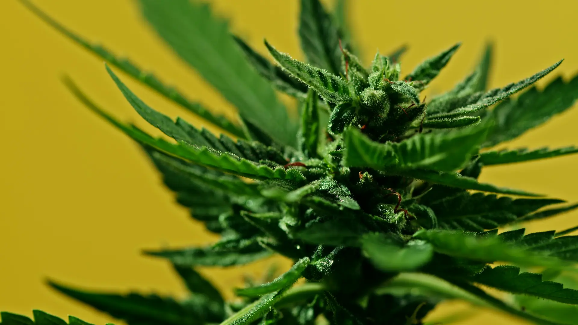
Root-zone waterlogging deprives root cortical cells of dissolved oxygen, shutting down ATP-driven ion transport and triggering widespread foliar chlorosis.
1. Overwatering and Root Hypoxia
When a container is irrigated too frequently without allowing sufficient gravitational dry-back, macropores remain saturated with liquid water. The oxygen diffusion rate in water is 10,000 times slower than in air. Within 24 to 36 hours of waterlogging, root cortical cells deplete dissolved oxygen, entering an anaerobic state (hypoxia).
Under hypoxic conditions, oxidative phosphorylation collapses; root cells run out of ATP, shutting down the active transmembrane proton pumps (H+-ATPase) required to absorb nitrate, potassium, calcium, and iron. Plants display widespread, droopy, generalized yellowing across both lower and middle foliage simultaneously. Inexperienced growers mistake this for nitrogen starvation and add more liquid fertilizer, further elevating root-zone salinity and accelerating root suffocation.
2. Substrate Core Temperature Extremes
Root metabolism is strictly temperature-dependent, following the biochemical Q10 temperature coefficient. When container substrate temperatures fall below 18 °C (64 °F) – common in winter grows where pots sit directly on cold concrete floors – root membrane fluidity decreases and enzymatic transporter activity drops by over 50%. Roots cannot assimilate phosphorus, magnesium, or iron, producing widespread chlorosis and purpling petioles regardless of fertilizer concentration.
Conversely, when root-zone temperatures exceed 26 °C (79 °F), dissolved oxygen solubility plummets while root respiration spikes, accelerating pathogenic colonization by waterborne oomycetes such as Pythium ultimum.
3. Atmospheric Vapor Pressure Deficit (VPD) Lockout
Transpirational mass flow in the xylem conduits pulls dissolved calcium and magnesium upward from the roots. If grow room air is excessively humid (VPD < 0.6 kPa), stomatal transpiration stops, stalling mineral transport. If the air is excessively dry (VPD > 1.6 kPa), stomatal guard cells close to prevent desiccation, stopping carbon fixation and inducing osmotic leaf tip burn and marginal chlorosis.
4. Mechanical Wind Burn and Fan Desiccation
High-velocity oscillating fans directed squarely at canopy foliage physically strip the protective boundary layer of humid air from the abaxial (underside) leaf surface. Stomata are forced into hyper-transpiration, losing water faster than root xylem conduits can resupply it.
The leaf responds defensively: outer margins curl downward into a claw shape, become rigid and brittle, and turn a pale, bleached yellow before developing papery brown necrotic patches. Wind burn chlorosis is strictly localized: leaves directly in the jet stream of the fan show severe symptoms, while leaves positioned just 20 cm below or behind the airstream exhibit pristine green morphology.
Grower Question: Responding to Sudden Lower Canopy Yellowing in Week 4
Why are our bottom fan leaves turning bright yellow and dropping in Week 4 of flower?
Question sent by: Darren Holbrook, via email.
“We are running a 9-week hybrid in 3-gallon coco coir pots under 900 μmol/m2/s LED fixtures. At Day 28 of flower, our bottom four fan leaves on almost every plant have turned completely pale yellow, withered, and fallen off when touched. The upper colas look dark green, healthy, and frosty. Our input feed is 2.2 dS/m at pH 5.8, but our runoff EC is sitting at 3.6 dS/m. Should we increase nitrogen to keep the bottom leaves green, or is this salt burn?”
Darren, your crop is experiencing a combination of natural light-driven source-sink reallocation aggravated by high root-zone pore-water salinity. Absolutely do not increase nitrogen.
In Week 4 of flowering, your canopy has developed a dense upper trellised ceiling. The bottom fan leaves near the substrate receive less than 100 μmol/m2/s PPFD – well below the photosynthetic light compensation point where carbon fixation equals respiratory consumption. Because these lower leaves are net carbon consumers, the plant actively triggers the PAO pathway, dismantling their chlorophyll and translocating their nitrogen and magnesium up into the rapidly swelling apical colas.
However, your leachate runoff EC of 3.6 dS/m (Delta EC +1.4 dS/m) indicates that unabsorbed fertilizer salts have accumulated in your coco pores, creating osmotic drought. This high salinity accelerates lower leaf senescence because the plant cannot maintain hydraulic turgor pressure in shaded tissue.
To stabilize your canopy and protect your colas, execute these two clinical actions:
- Execute a Stepped Reset Fertigation: Mix a complete floral baseline solution at a reduced input EC of 1.4 to 1.5 dS/m buffered to pH 5.8. Irrigate thoroughly with a 35% to 40% leaching fraction. This sweeps the concentrated salt crusts out of the container base without shocking root hairs. Once runoff drops below 2.5 dS/m, resume feeding at an adjusted input EC of 1.9 dS/m with a consistent 20% daily leaching fraction.
- Prune the Shaded Under-Canopy: Perform a clean bottom lollipop pruning: remove the yellowing, shaded lower leaves and underdeveloped micro-suckers from the bottom third of the plant. This improves under-bench airflow, eliminates potential vectors for powdery mildew and Botrytis, and allows the plant to focus 100% of its transpirational energy on developing dense, frosty top colas.
The Complete Cannabis Foliar Chlorosis Diagnostic Matrix
The following empirical matrix differentiates every major physiological, chemical, and biological cause of leaf yellowing in cannabis:
| Primary Root Cause | Canopy Location | Visual Pattern & Progression | Key Environmental / Chemical Trigger | Immediate Corrective Action |
|---|---|---|---|---|
| Nitrogen (N) Deficiency | Oldest bottom fan leaves first, advancing upward. | Uniform pale yellowing across entire leaf, including veins; leaves wilt soft and drop cleanly. | Input EC < 1.2 dS/m in vegetative growth; depleted organic substrate. | Increase nitrate nitrogen input to 160-180 ppm N; check Delta EC. |
| Magnesium (Mg) Deficiency | Lower to mid-canopy mature leaves. | Sharp interveinal chlorosis (green veins, yellow lamina) with rusty necrotic flecks along margins. | Potassium excess (K > 250 ppm) or substrate pH < 5.5. | Lower input potassium; foliar spray 1% Epsom salt; reset substrate pH to 5.9. |
| Iron (Fe) Lockout | Top apical growing tips and new sugar leaves. | Bright lemon-yellow to white interveinal bleaching; veins remain dark green. | Substrate pH > 6.4 in soilless; cold wet media (<18 °C). | Switch to Iron DTPA / EDDHA; reset substrate pH to 5.7 with stepped leach. |
| Root Hypoxia (Overwatering) | Generalized; affects lower and middle canopy simultaneously. | Leaves turn pale yellow, droop heavy with turgid water-swollen petioles; firm curl downward. | Irrigating before achieving 15% to 20% VWC dry-back; poor container drainage. | Extend dry-back interval; ensure drainage holes are unobstructed; aerate room. |
| Apical Photobleaching | Terminal crowns directly below LED fixtures only. | Both veins and lamina turn chalk-white; leaves 15 cm lower remain completely green. | PPFD > 1,100 μmol/m2/s without supplemental CO2. | Dim LED lights by 20% or raise fixtures 20 cm; elevate CO2 to 1,200 ppm. |
| Natural Late-Flower Fade | Broad canopy fan leaves during final 14 days before harvest. | Gradual mosaic yellowing interspersed with anthocyanin purples, reds, and golds. | Normal senescence; nitrogen withdrawal; trichome amber maturation. | Do nothing; maintain harvest schedule; celebrate healthy terpene expression. |
Common Mistakes, Look-Alikes, and Dangerous Rules of Thumb
Navigating foliar yellowing requires avoiding unscientific cultivation myths that turn minor anomalies into harvest-destroying crises:
- Myth 1: “All yellow leaves need nitrogen.” Applying high-nitrogen fertilizers when the true cause is root hypoxia or high substrate salinity adds toxic mineral solute mass to suffocating root hairs. The result is rapid root tip necrosis, leaf clawing, and accelerated leaf drop.
- Myth 2: “Flush with pure reverse osmosis water to clear yellowing.” Flushing saturated media with pure 0.0 dS/m water causes violent osmotic shock. Water rushes into root cells via hypoosmotic pressure, bursting fine root hairs and stripping the calcium-magnesium saturation buffer from coco coir fibers. Always use stepped, reduced-EC complete nutrition (1.2 to 1.4 dS/m) for root-zone resets.
- Myth 3: “Strip off all yellow leaves immediately.” While pruning severely diseased or shaded lower suckers is beneficial, ripping off slightly chlorotic fan leaves in mid-flower removes the plant’s emergency mobile nutrient reserves. If the root zone remains deficient, the plant will simply cannibalize the next layer of healthy leaves above them. Fix the root cause first before defoliating.
Microscopic Pest and Viral Look-Alikes
Two non-nutritional biological agents routinely mimic severe nutrient chlorosis:
- Broad Mites and Russet Mites: These microscopic arachnids (<0.2 mm) are invisible to the naked eye. As they feed on tender epidermal cells of apical new growth, they inject toxic saliva containing digestive enzymes. Emerging leaves turn bronze-yellow, curl rigidly downward like plastic, and develop glossy or glassy textures commonly misdiagnosed as extreme nitrogen toxicity, boron deficiency, or over-fertilization. Always inspect the undersides of yellow curled leaves with a 60x to 100x microscope.
- Hop Latent Viroid (HLVd) and Tobacco Mosaic Virus (TMV): Systemic plant pathogens alter nuclear gene expression and chloroplast division. Infected plants display irregular, asymmetric yellow mosaic blotching, horizontal leaf twisting, brittle main branches, and severe floral stunting (“duding”). If foliar yellowing exhibits random blotchy marbling rather than symmetrical vascular patterns, submit leaf tissue for RT-PCR laboratory testing.
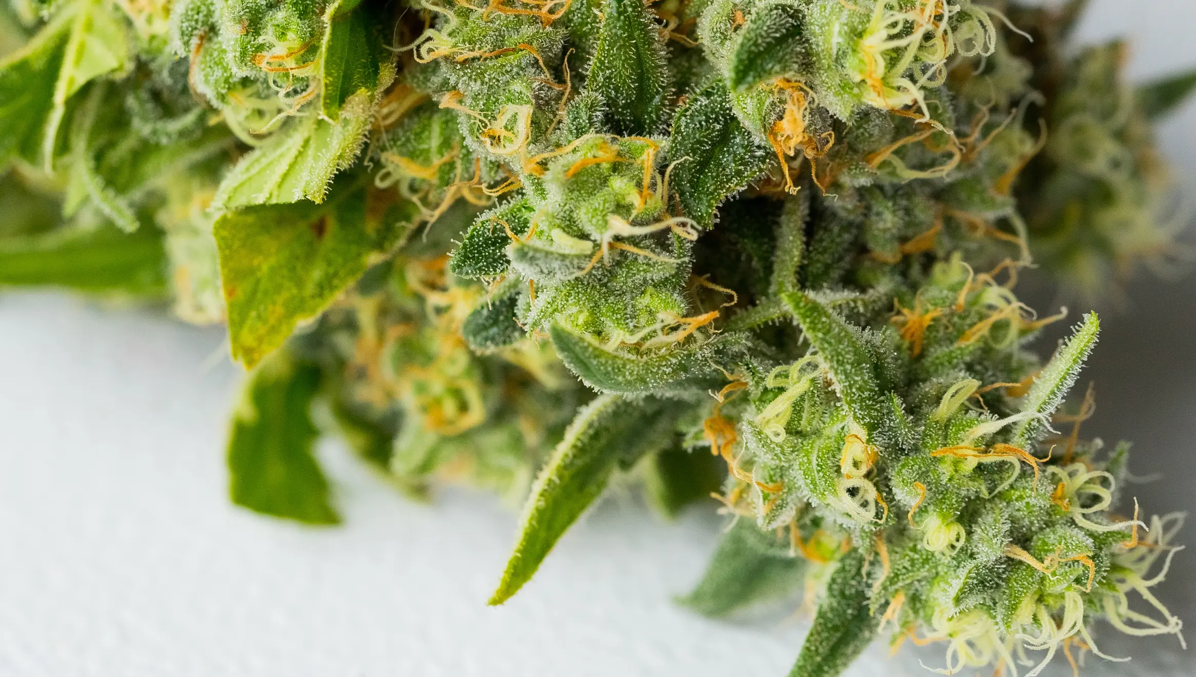
Late-stage floral senescence represents a natural programmed remobilization of carbohydrates, distinct from premature pathogen-induced chlorosis.
How to Verify Root Cause: PourThru Extraction, Delta EC, Petiole Sap, and Tissue Testing
Never implement corrective interventions based on visual scouting alone. Verify root-zone and canopy status through four objective, repeatable measurements:
1. The PourThru Pore-Water Extraction
Standard tray runoff degrades through atmospheric CO2 outgassing within minutes, distorting pH readings. Perform a standardized PourThru test (Cavins et al., 2000): one hour following an irrigation event, pour 200 mL of distilled water onto the surface of a 3-gallon pot. Collect and measure the initial 50 mL of displaced leachate. If PourThru pH is above 6.4, yellowing is driven by iron/manganese lockout. If PourThru pH is below 5.5, yellowing is driven by magnesium/calcium lockout.
2. Delta EC (ΔEC) Salinity Audit
Measure input feed EC versus composite runoff EC (ΔEC = ECout – ECin). If Delta EC exceeds +0.8 dS/m, high osmotic pressure is preventing water and nutrient uptake, causing induced foliar chlorosis. A stepped volumetric leaching reset is required.
3. Handheld Petiole Sap Analysis and Chlorophyll Fluorometry
For real-time operational feedback without waiting 5 to 7 business days for laboratory mail-in results, commercial facilities utilize handheld ion-selective electrode (ISE) sap meters (such as Horiba LAQUAtwin) and chlorophyll fluorometers:
- Petiole Sap Nitrate (NO3–): Collect 5 petioles from recently matured fan leaves, extract sap using a hydraulic garlic press, and test immediately. Vegetative cannabis should read 800 to 1,200 ppm NO3–. Readings below 500 ppm confirm true systemic nitrogen exhaustion, while readings above 1,000 ppm in yellowing plants prove root-zone lockout or hypoxia rather than fertilizer scarcity.
- Chlorophyll Fluorometry (Fv/Fm Ratio): Dark-adapt target leaves for 20 minutes with leaf clips before measuring Photosystem II maximum photochemical quantum efficiency. Healthy cannabis exhibits an Fv/Fm ratio between 0.80 and 0.84. A sharp drop below 0.72 indicates acute photoinhibition, light stress, or severe oxidative damage to thylakoid reaction centers.
- SPAD Optical Chlorophyll Index: Handheld SPAD-502 meters measure relative foliar chlorophyll density non-destructively. Values dropping below 38 SPAD units signify active chlorophyll catabolism before visible leaf chlorosis is discernible to the naked eye.
4. Laboratory Leaf Tissue Analysis (ICP-OES / Dumas)
Harvest the Most Recently Matured Leaf (MRML) at node 4 or 5 and submit to a certified agronomic testing laboratory. Total nitrogen below 3.5% dry weight in vegetative growth confirms genuine nitrogen starvation. Leaf iron below 60 mg/kg Fe confirms iron deficiency.
Systematic Recovery SOP: The Single-Variable Correction Sequence
When foliar yellowing emerges, follow this disciplined single-variable elimination workflow:
Phase 1: Environmental and Moisture Verification
Measure canopy PPFD with a quantum sensor. Verify lights-on room temperature is 26 °C to 28 °C and room VPD is 1.0 to 1.3 kPa. Next, probe substrate core temperature: verify it operates between 20 °C and 23 °C. Check container moisture weight: if the pot is heavy and waterlogged, delay the next feeding event until 18% to 22% volumetric water content dry-back is achieved.
Phase 2: The Root-Zone Chemical Reset
If PourThru pH is outside 5.8 to 6.2 or Delta EC exceeds +0.8 dS/m, execute a stepped reset fertigation: formulate a complete baseline nutrient solution at 1.4 dS/m EC buffered to pH 5.7 (if substrate is alkaline) or pH 6.1 (if substrate is acidic). Administer a generous 35% to 40% leaching fraction to displace accumulated salts and re-establish chemical equilibrium.
Phase 3: Emergency Foliar Rescue Applications
Because damaged or waterlogged roots require 48 to 72 hours to re-establish normal hydraulic and active ion uptake, foliar feeding offers an immediate trans-cuticular bypass:
- For Mobile Deficiencies (N, Mg): Apply pharmaceutical-grade magnesium sulfate (Epsom salt) at 5 g/L (0.5% w/v) combined with amino-acid chelated nitrogen at 0.5 mL/L. Ensure spray water is buffered to pH 6.2 with a non-ionic organosilicone surfactant. Apply right as lights dim to prevent lens scorching.
- For Immobile Deficiencies (Fe, Zn, Mn): Apply iron chelate (Fe-DTPA or Fe-EDDHA) at 0.5 to 1.0 g/L. Cuticular absorption of iron directly stimulates localized chlorophyll re-synthesis within 24 to 36 hours while root-zone substrate pH is being corrected.
Phase 4: Observation and Verification
Change only one variable at a time. After executing the reset, maintain stable fertigation and monitor the newest emerging apical growth. Yellowed lower leaves will rarely turn fully green again, but the progression of yellowing should halt completely within 48 to 72 hours, and all new leaves should emerge dark emerald green.
Day-by-Day Physiological Recovery Timeline
Understanding the internal biological timeline prevents growers from panicking and stacking unnecessary corrections:
- Hours 0 to 24 (Osmotic Equilibration): The reduced-salinity reset dissolves crystallized mineral crusts in the substrate matrix. Root cortical cells regain osmotic turgor pressure; stomatal conductance increases from near-zero to 40% of baseline. Leaf petiole angles rise as hydraulic pressure is restored.
- Days 2 to 3 (Enzymatic Downregulation of Catabolism): As newly available ionic nitrate, iron, and magnesium flood the vascular xylem stream, nuclear genes down-regulate pheophorbide a oxygenase (PAO) and chlorophyllase enzymes. Chlorophyll catabolism halts in newly expanding leaves.
- Days 4 to 7 (Chloroplast De Novo Assembly): Newly emerging shoot apical meristems and sugar leaves synthesize fresh thylakoid membranes and light-harvesting complexes. Emerging foliage displays a pristine, dark emerald green color. While severely yellowed lower fan leaves will not regain green pigmentation (their chloroplasts having degraded into irreversible gerontoplasts), their yellowing progression halts entirely.
- Days 7 to 10 (Restoration of Maximum Net Carbon Fixation): Whole-canopy net photosynthetic rate (A_net) returns to maximum capacity (typically 25 to 35 μmol CO2/m2/s under high PPFD). Vegetative biomass accumulation and floral bulking resume full velocity.
Canopy Health Standard Operating Inspection Checklist
Enforce these standardized diagnostic checkpoints across every crop scouting walk:
- Canopy location of yellowing is mapped: lower leaves (mobile N/Mg) vs upper meristem tips (immobile Fe/S/Zn).
- Container weight and dry-back are audited before diagnosing nutrient deficiency to rule out root hypoxia.
- Substrate root-zone temperature is confirmed between 20 °C and 23 °C using a digital soil probe.
- PourThru pore-water extraction is conducted; substrate pH is maintained strictly between 5.8 and 6.2.
- Delta EC is monitored daily; runoff EC exceeding input by +0.8 dS/m triggers an immediate stepped leaching reset.
- Apical crowns directly under LED hotspots are checked for photobleaching before altering fertilizer recipes.
- Pure water flushing is strictly avoided; salinity resets utilize stepped 1.4 dS/m balanced nutrition.
- Late-flower natural fade (Weeks 7-9) is recognized as normal senescence and protected from panic feeding.
- Bottom lollipop pruning is performed on severely shaded under-canopy leaves to optimize airflow.
- Only one environmental or nutritional variable is adjusted at a time, followed by a 72-hour recovery audit.
Scientific References
- Barker, A. V., & Pilbeam, D. J. (2015). Handbook of Plant Nutrition (2nd ed.). CRC Press, Boca Raton, FL.
- Bernstein, N., Gorelick, J., Zerahia, R., & Koch, S. (2019). Impact of environmental conditions and nutrient availability on secondary metabolite production in medical cannabis. Industrial Crops and Products, 138, 111467.
- Cavins, T. J., Whipker, B. E., Fonteno, W. C., Harden, B., McCall, I., & Gibson, J. L. (2000). Monitoring and Managing pH and EC Using the PourThru Extraction Method. Horticultural Information Leaflet 590, North Carolina State University.
- Hörtensteiner, S., & Kräutler, B. (2011). Chlorophyll breakdown in higher plants. Biochimica et Biophysica Acta (BBA) – Bioenergetics, 1807(8), 977-988.
- Marschner, H. (2012). Marschner’s Mineral Nutrition of Higher Plants (3rd ed.). Academic Press, London.
- Taiz, L., Zeiger, E., Møller, I. M., & Murphy, A. (2015). Plant Physiology and Development (6th ed.). Sinauer Associates, Sunderland, MA.
- Zheng, Y. (2021). Handbook of Cannabis Production in Controlled Environments. CRC Press, Boca Raton, FL.
Share this article
A quick overview of the topics covered in this article.