
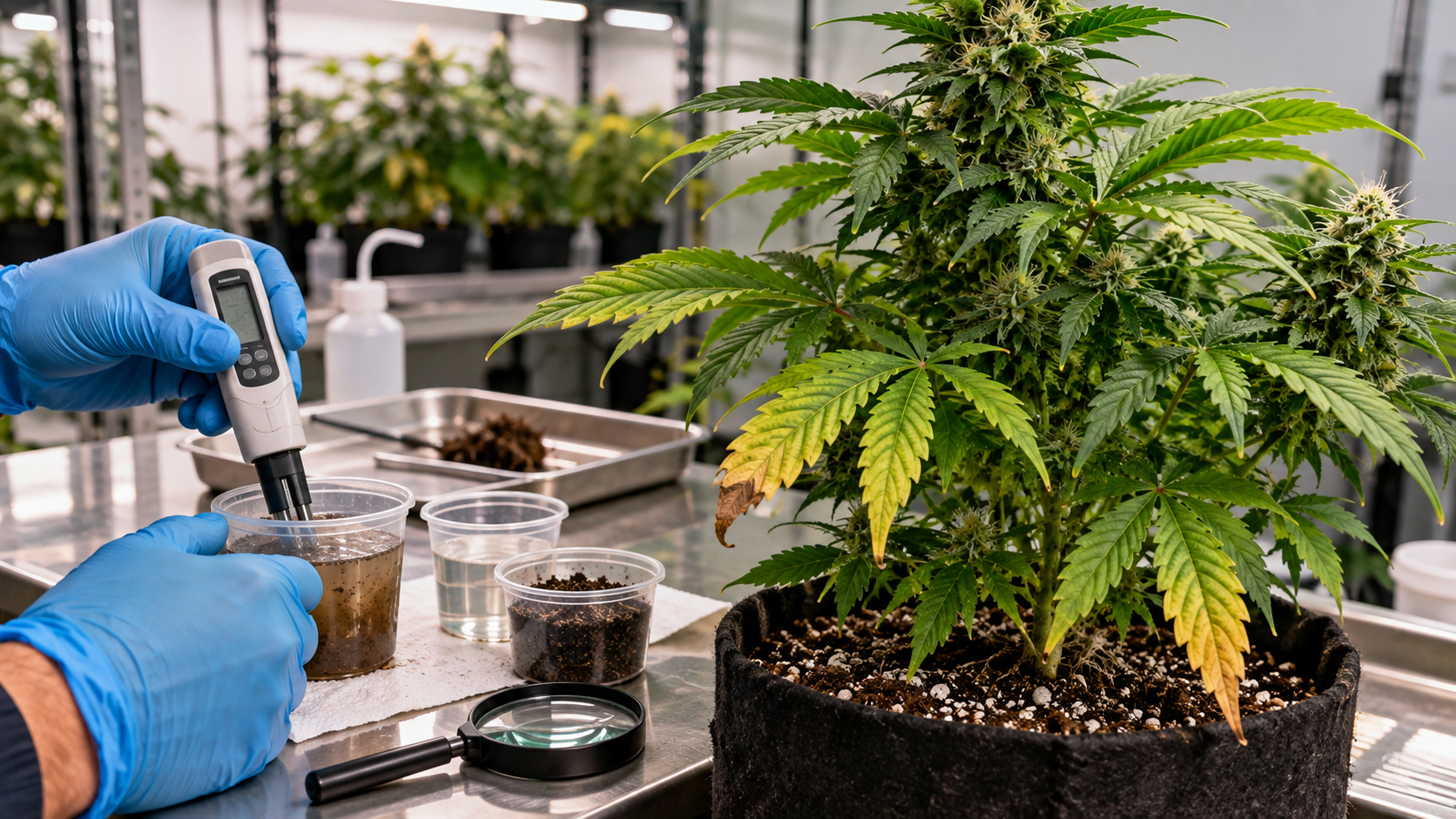
Fusarium on Cannabis: Wilt, Root Rot, Crown Rot, and Stem Canker
Fusarium on cannabis is not one disease with one unmistakable appearance. Different pathogenic Fusarium species and isolates can be associated with damping-off, root and crown rot, vascular wilt, pith necrosis, stem cankers, and flower disease. The same genus also includes organisms that may be recovered from a plant without proving that they caused its decline.
This is why a wilted branch, a brown stem center, or discolored roots should begin an investigation rather than end it. Water stress, root-zone oxygen problems, salt injury, physical damage, Pythium, Diaporthe, Sclerotinia, insects, and other causes can overlap with a Fusarium-compatible pattern.
The useful workflow is to map where the problem appears, inspect the roots, crown, vascular tissue, pith, and outer stem as appropriate, preserve an untreated sample, contain possible movement, and obtain a laboratory diagnosis when the answer will affect stock plants, crop removal, water treatment, or facility sanitation.
What Fusarium Disease Means on Cannabis
Fusarium is a large genus of fungi, not a single cannabis pathogen. Cannabis research has reported numerous Fusarium species in association with diseased plants, but association is not the same as demonstrated pathogenicity. Some isolates have been tested and shown to cause disease; others have only been detected or recovered. Species identity, isolate behavior, plant tissue, crop stage, and growing conditions all affect what a result means.
On cannabis, pathogenic isolates can attack different tissues. Seedlings or unrooted cuttings may damp off. Roots and the crown may rot. Vascular tissue can discolor and lose function, producing wilt. The pith may become necrotic. An external stem lesion may expand into a canker, and some Fusarium species have also been associated with flowers. A diagnosis should therefore name both the organism and the disease form when the evidence allows it. Use the Cannabis Damping-Off guide when the failure occurs before emergence or during the seedling and cutting stage.
Fusarium disease
Root, crown, vascular, stem, pith, propagation, or flower disease caused by a pathogenic Fusarium isolate. The disease form and organism must be supported by affected tissue, crop pattern, growing history, and appropriate laboratory evidence.
A field study of cannabis plants with yellowing, wilting, and collapse recovered Fusarium species and Pythium aphanidermatum from root and crown disease. Other cannabis studies have documented Fusarium in stock plants, cuttings, propagation areas, drainage water, stems, and pith. Together, these findings support a broad disease complex. They do not support calling every wilt or brown root Fusarium.
Important: A laboratory detection and a disease diagnosis are related but different. Detection shows that an organism was found in the submitted material. Diagnosis asks whether that organism, in that tissue and crop pattern, explains the observed damage.
Why the disease form matters
A localized stem canker on one wounded branch creates a different decision from progressive crown rot in a mother plant or root disease moving through a recirculating system. The first may be associated with one injury site. The others may threaten every cutting or plant connected to the same stock or water path.
Describe what is actually affected before choosing a response. Useful terms include root rot, crown rot, vascular discoloration, wilt, pith necrosis, stem canker, and damping-off. Avoid using “Fusarium wilt” as a catch-all for every disease involving this genus.
Predisposing stress is not proof of infection
Root injury, transplant damage, pest feeding, prolonged wetness, poor drainage, oxygen stress, high soluble salts, and stem wounds can increase vulnerability or help an existing infection progress. In cannabis pathogenicity research, wounding was important for some Fusarium isolates to produce severe crown and stem symptoms under the study conditions.
This does not mean a wound automatically becomes infected or that every stressed plant contains Fusarium. Disease still requires a pathogenic organism, a susceptible host, a suitable infection court, and conditions that allow colonization.
How to Identify a Fusarium-Compatible Pattern
Begin with the pattern across the grow. Record whether decline follows a mother plant, cutting batch, cultivar, bench, water loop, transplant date, wound type, low area, or hot period. Compare suspect plants with healthy plants of the same age and management history. A repeated pattern carries more diagnostic weight than one isolated symptom.
Canopy symptoms point below the surface
Possible aboveground signs include stunting, loss of vigor, yellowing, reduced water use, midday wilt, wilt that does not resolve normally, one-sided branch decline, premature leaf loss, and collapse. Plants with damaged roots or vascular tissue may wilt during the greatest evaporative demand before they remain wilted throughout the day.
These symptoms indicate that water uptake or transport is failing. They do not reveal why. Check irrigation delivery, medium moisture, root integrity, crown condition, environmental demand, and mechanical damage before assigning an organism name.
Roots and crown provide stronger evidence
Fusarium-compatible root disease may include reduced fine roots, brown or black lesions, sunken tissue, decay, and a smaller functional root system. The crown may show an external sunken lesion or internal brown discoloration extending upward from the root zone. Tissue can be firm in some vascular or canker cases and soft when decay is advanced or mixed organisms are present.
Color alone is weak evidence. Nutrient solutions, organic inputs, media particles, root age, oxygen stress, salts, and other pathogens can all change root color. Compare tissue texture and distribution, not just hue. A sharp transition between healthy and damaged tissue is useful sampling material.
Internal stem symptoms require careful interpretation
Brown vascular streaking, a discolored crown, or necrotic pith may support a vascular, crown, or stem disease investigation. None is unique to Fusarium. Cutting a living plant is destructive, creates a new wound, and can spread contaminated sap or tissue on the blade. Section only material already selected for removal or laboratory submission unless a qualified diagnostician directs otherwise.
Warning: Do not split valuable stock plants open merely to look for a brown stem center. Photograph external symptoms, preserve representative material, and contact the diagnostic laboratory before collecting a destructive sample.
External stem cankers may carry visible clues
A canker is a localized area of dead bark or stem tissue. It may appear sunken, dry, cracked, discolored, or bordered by callus. The stem above an expanding lesion may wilt if water-conducting tissue is interrupted. Under favorable conditions, some fungi form visible structures or spores on dead tissue, but absence of visible growth does not exclude disease.
Record whether the lesion begins at a pruning cut, tie, trellis rub, broken branch, insect injury, soil line, or splash zone. That location can explain the infection court and help distinguish a wound-associated canker from disease moving upward from roots and crown.
Grower question: One branch is wilting and the pith looks brown. Does that prove Fusarium?
No. The pattern is compatible with several vascular, pith, canker, borer, and mechanical problems. Inspect the outside of the stem, the crown, roots, irrigation path, wounds, and possible insect galleries. Submit the transition between healthy and damaged tissue when confirmation will change what you remove or sanitize.
Question sent by: J_Morrison, via email.
Use a laboratory when the decision is consequential
Contact the laboratory before sampling. Depending on the case, it may request intact roots, crown sections, the advancing edge of a stem lesion, or several plants at different symptom stages. Include a healthy comparison when possible. Keep roots and lower stems associated with the same plant, and prevent wet tissue from decomposing in a sealed, overheated package.
Diagnostic work may combine visual examination, fungal isolation, microscopy, pathogenicity information, and molecular identification. Sequencing regions such as translation elongation factor 1-alpha can help distinguish Fusarium species more effectively than appearance alone. Ask what the method can identify, whether mixed infections were assessed, and how the result fits the submitted tissue.
Field Advice: Preserve an untreated sample before applying fungicides, oxidizers, disinfectants, biological products, or home remedies. A panic treatment can reduce the chance of recovering the original organism without repairing diseased tissue.
Fusarium Look-Alikes
Fusarium symptoms overlap with infectious and noninfectious problems. A useful differential diagnosis does not produce a longer guess list. It identifies the next observation or test that can separate the leading possibilities.
| What you find | Possible explanation | Best next check |
|---|---|---|
| Wet medium, wilt, roots mostly firm, no expanding lesion | Overwatering or low root-zone oxygen without confirmed infection | Verify moisture at depth, drainage, root texture, water use, and response after the physical error is corrected |
| Soft roots with cortex slipping away and rapid loss of fine roots | Pythium, another oomycete, Fusarium, or mixed root rot | Contain shared water and submit roots plus crown tissue using the laboratory’s protocol |
| Firm crown or vascular browning with progressive wilt | Fusarium, Verticillium, bacterial disease, or another vascular and crown disorder | Sample the healthy-to-damaged transition and request organism-level interpretation |
| Dry external canker beginning at a wound | Fusarium, Diaporthe, Botryosphaeriaceae, mechanical damage, or another canker pathogen | Inspect lesion margins and fruiting structures; submit the advancing edge rather than dead center tissue alone |
| Cottony growth, water-soaked stem base, or hardened resting bodies | Sclerotinia, southern blight, or another stem and crown rot | Preserve visible structures and submit intact tissue without cleaning them away |
| Hole, frass, hollowing, or a gallery in the stem | Borer injury, sometimes followed by secondary colonization | Open removed material over a clean surface and identify the insect or gallery pattern |
| Burned root tips, high verified root-zone conductivity, surviving roots remain firm | Salt or fertilizer injury | Verify input and root-zone measurements with a method appropriate to the medium |
| Localized damage after transplant, staking, pruning, or tie pressure | Mechanical injury with or without secondary infection | Trace the damaged area to the handling event and watch whether the margin continues to expand |
Pythium can overlap with Fusarium
Both groups can cause root and crown disease, stunting, yellowing, and wilt. Pythium-compatible roots often become water-soaked and lose their outer cortex, but Fusarium and mixed infections can also produce substantial decay. A shared reservoir strengthens concern about a waterborne pathway without naming the organism.
Use Pythium Root Rot on Cannabis when soft root decay and a connected water system are central to the case. Use Root Rot vs. Overwatering when the first question is whether the root zone is physically too wet or actively decaying.
Canker pathogens can look alike
A stem lesion should not be named from color or shape alone. Several fungi can colonize wounded cannabis stems, and dead tissue may later carry secondary organisms. Sample the advancing lesion margin, where living and damaged tissues meet. The dry center of an old canker may no longer contain the primary pathogen in a useful form.
Water stress can imitate vascular disease
A blocked emitter, damaged root ball, failed pump, root-bound container, extreme evaporative demand, or broken stem can create sudden wilt. Confirm that water reaches the root zone and that the plant can take it up. Adding more water without checking moisture can worsen oxygen stress and root disease.
Do: Combine crop pattern, tissue location, root and crown integrity, wound history, water pathways, progression, and appropriate testing.
Avoid: Diagnosing Fusarium from a single wilted branch, brown stem center, or discolored root system.
How Fusarium Enters and Moves Through a Grow
Fusarium can enter production with infected stock plants, cuttings, seedlings, seed-associated material, contaminated media or soil, irrigation water, equipment, footwear, debris, and transplants. The important pathway differs by facility. Trace actual connections instead of assuming that every case arrived from the same source.
Mother plants and cuttings can connect crop generations
Vegetative propagation preserves a desirable genotype, but it can also preserve and distribute a pathogen. Cannabis research has recovered Fusarium oxysporum from symptomatic stock plants, associated propagation areas, and a small proportion of visually asymptomatic cuttings under the study conditions. This supports a strict lesson: appearance alone cannot certify a cutting as clean.
If several young plants from one mother decline, stop taking more cuttings from that stock line while the cause is investigated. Keep source records that connect every tray to its mother, propagation date, worker or station, rooting input, and destination. Traceability can turn a facility-wide mystery into a defined batch decision.
Water and drainage may connect plants
Fusarium propagules have been recovered from drainage and production water in cannabis facilities. Shared reservoirs, flood tables, runoff channels, hoses, tanks, filters, and recirculating plumbing can therefore be part of the movement pathway. Water does not need to look dirty to carry biological risk.
Map the direction of flow and every point where clean water can contact runoff, roots, floor splash, or debris. In drain-to-waste production, confirm that one container cannot drain into another. In recirculating production, treat the connected water system as part of the investigation rather than cleaning only the visibly sick plant.
Wounds create entry points
Transplant tears, pruning cuts, trellis abrasion, tight ties, broken branches, insect feeding, and rodent damage can expose susceptible tissue. In cannabis field research, crown and stem disease developed after wound inoculation with selected Fusarium isolates. Sporulation on diseased tissue can also add inoculum to the immediate environment.
Reduce avoidable injury and clean tools between plants. Do not assume that a clean pruning cut prevents all disease, but recognize that contaminated blades can combine wounding and inoculum placement in one action.
Soil and debris can preserve inoculum
Many Fusarium species can survive in soil, plant residues, roots, or resistant structures. Removing the canopy while leaving infected roots, fallen tissue, dirty stakes, contaminated media, or inaccessible debris can allow the same pattern to return. Broad host ranges also limit the value of casual crop rotation as a stand-alone solution.
What to Do When Fusarium Is Suspected
The first response should protect healthy plants and preserve evidence. Do not begin by applying several products to the whole grow. Contain possible movement, document the untreated pattern, correct verified physical failures, and decide what needs laboratory confirmation.
1. Stop propagation and shared movement
Do not take more cuttings from a suspect mother. Separate new material from established clean stock. Prevent runoff, tools, gloves, carts, stakes, ties, media, and plant debris from moving from suspect plants into clean production. Work clean areas first and the affected zone last.
If plants share water, identify the full connected group. Isolate a unit only when the system design permits it without creating an irrigation, electrical, pressure, or oxygen failure. A connected reservoir can make apparently healthy plants part of the same exposure event.
2. Document the crop pattern
Photograph the room, bench, whole plant, crown, roots, external stem lesion, and a healthy comparison. Record the source plant, cultivar, propagation and transplant dates, irrigation history, water source, shared equipment, recent pruning, wounds, pest activity, environmental events, and every product already applied.
Mark affected plants on a simple map. Note whether the pattern follows one mother, one worker or tool sequence, one water line, the wettest area, a wound type, or a plant age. Include symptom-free exposed plants in the map.
3. Preserve representative samples
Call the plant diagnostic laboratory before cutting. A useful sample often includes roots, crown, and a stem section spanning the transition from apparently healthy to affected tissue. A canker sample should include the active margin and surrounding tissue. Submit more than a dry dead center whenever the laboratory advises it.
Keep samples labeled by plant and location. Do not wash away spores, surface structures, root fragments, or lesion margins unless instructed. Package and ship according to the laboratory’s requirements, because overheated or decomposed tissue can obscure the primary problem.
4. Correct confirmed physical failures
Restore a failed emitter, blocked drain, pump, aeration device, or circulation component using the safe procedure for that system. Stop standing runoff and unnecessary irrigation. Remove tie pressure or continuing abrasion. These corrections support remaining healthy tissue but do not disinfect established disease.
5. Define removal boundaries
Remove collapsed plants and loose infected debris in a way that contains roots and media. Do not drag them through clean production. For valuable plants with localized symptoms, base the hold-or-remove decision on lesion location, progression, laboratory findings, stock value, proximity to other plants, and whether the plant is serving as propagation material.
Warning: Cutting a canker out of a plant does not prove the infection is gone. The organism may extend beyond the visible margin or already involve vascular, crown, root, or connected propagation tissue.
Grower question: Can I cut the damaged section out and keep the plant?
Sometimes a truly localized wound can be removed, but appearance alone cannot define the infection boundary. A canker near the crown, systemic wilt, multiple lesions, root involvement, or disease in a mother plant creates a much higher consequence. Isolate the plant, preserve a sample, and let lesion progression and diagnostic evidence guide the decision.
Question sent by: GreenhouseGary, via contact form.
Soil, Coco, Hydroponic, and Stock-Plant Response
The organism does not change because the growing medium changes, but the exposure pathways, useful measurements, and containment options do. Use system-specific evidence rather than applying one generic dry-back or reservoir recipe.
| Production system | Inspect first | Containment priority | Common mistake |
|---|---|---|---|
| Mother and propagation stock | Source-line pattern, crown, pruning sites, rooting success, tray and tool history | Stop cuttings from suspect stock and trace every distributed batch | Keeping an asymptomatic cutting as a replacement without quarantine or testing |
| Soil or peat containers | Roots, crown, moisture at depth, drainage, transplant injury, runoff contact | Contain roots, media, runoff, stakes, and tools by plant or zone | Watering again because vascular wilt resembles drought |
| Coco or other soilless substrate | Emitter function, root-zone measurements, plug-to-media transition, roots and crown | Trace the irrigation batch and prevent return-flow or drain contamination | Blaming conductivity alone while a crown lesion continues to expand |
| Recirculating hydroponics | Root integrity, reservoir, return lines, filters, biofilm, water source, connected plants | Include the full water loop in sampling and sanitation planning | Removing one plant while leaving exposed water and plumbing unexamined |
| Outdoor soil or beds | Low areas, drainage, crown injury, field equipment, rodents, prior crop and residue | Limit movement of wet soil, roots, debris, and contaminated equipment | Relying on a short rotation without knowing the Fusarium species or host range |
Mother plants require the strictest threshold
A mother plant is not just one plant. It is a distribution point for many future plants. Recurring wilt, crown discoloration, unexplained cutting loss, or confirmed Fusarium around stock deserves a lower tolerance than a non-propagated plant near harvest. Quarantine suspect stock, stop distribution, and test material selected with the laboratory.
Grower question: A clone looks clean even though its mother is suspect. Is it safe to keep?
Visual cleanliness does not establish that a cutting is pathogen-free. Keep it out of clean stock and shared water, maintain its source identity, and use an appropriate testing and observation plan. Do not allow one apparently healthy replacement to bypass the same investigation applied to its mother.
Question sent by: NorthernMike, via Facebook page.
Containers can hide crown disease
In soil, peat, or coco, the crown may be obscured by media and the root ball may stay wet at depth while the surface looks dry. Inspect the actual root zone and verify irrigation distribution. Avoid repeated removal and repotting of a suspect plant, which can spread media and create more wounds.
Correct drainage and irrigation errors, but do not interpret temporary canopy improvement as proof that a pathogen has been eliminated. Track lesion margins, new root growth, water use, and neighboring plants.
Recirculating water expands the unit of concern
In deep-water culture, nutrient film, flood-and-drain, or another recirculating design, the epidemiological unit may be the entire loop. Sample selection should represent both diseased tissue and the connected system. Filters, pumps, chillers, manifolds, return channels, inaccessible biofilm, and dead zones all matter when planning cleanup.
Water-treatment equipment must be sized and validated for the actual flow, water chemistry, organic load, organism, and crop system. A technology that reduces inoculum in one study is not automatically an effective or crop-safe setting for every facility.
Control Options and Their Limits
There is no universal product that reverses every Fusarium disease form. Necrotic roots, destroyed vascular tissue, and dead pith do not become healthy again. A successful program reduces new infection, contains movement, corrects predisposing conditions, protects clean material, and verifies that disease is no longer progressing.
Sanitation depends on complete removal
Remove diseased plants, roots, media, fallen tissue, and disposable supports according to local requirements. Clean visible organic matter before applying an appropriate disinfectant to compatible hard surfaces. Organic debris can shield organisms and consume disinfectant activity. Follow the product label for surface preparation, concentration, contact time, ventilation, personal protection, rinsing, and crop compatibility.
Some components cannot be cleaned reliably because they are porous, cracked, heavily biofilmed, or inaccessible. Replace them when the sanitation plan cannot reach or validate the contaminated surface. Keep clean tools separate from tools used during removal.
Fungicide use is label- and jurisdiction-specific
A Fusarium isolate being sensitive to an active ingredient in a laboratory does not mean the product is registered, effective, or safe for cannabis in a particular location. Crop stage, application site, residue rules, worker protection, resistance management, and local regulations all matter.
Use only products permitted for the crop, site, disease, and jurisdiction, exactly as the label directs. Do not extrapolate a root drench, seed treatment, or ornamental-crop result into a cannabis flower treatment. Where no appropriate product is registered, exclusion, removal, sanitation, and environmental correction carry even more importance.
Biological products are not a rescue guarantee
Some beneficial microbes and biological products may suppress Fusarium under particular experimental or production conditions. Their performance depends on strain identity, viability, timing, root-zone environment, compatibility with other inputs, and disease pressure. A preventive result does not demonstrate that a collapsed plant with crown and vascular destruction can be rescued.
If a biological is legally permitted and compatible with the system, evaluate it as one part of an integrated program. Preserve untreated diagnostic evidence first and use a defined comparison rather than adding several organisms and products at once.
Water treatment requires engineering, not guesswork
Filtration, heat, ultraviolet systems, ozone, oxidants, and other treatments differ in what they remove or inactivate. Their performance changes with flow rate, dose delivery, turbidity, organic matter, water chemistry, contact design, and maintenance. Some can harm roots, workers, equipment, or beneficial organisms when misapplied.
Use qualified guidance to design and validate water treatment. Measure the relevant operating parameters and confirm biological performance with an appropriate monitoring plan. Do not rely on reservoir clarity, odor, or a device’s power rating as proof of pathogen control.
Do: Build control around exclusion, containment, removal, clean stock, clean water pathways, sanitation, wound reduction, and verification.
Avoid: Combining unverified drenches or raising product strength because a wilted plant did not recover immediately.
Prevention and Verification
Prevention begins before symptoms appear. The strongest program limits entry, separates incoming material, protects mother stock, controls water and debris movement, reduces wounds, records plant lineage, and defines what evidence is required before a cleaned area returns to normal production.
Build a clean-stock boundary
Inspect incoming plants and keep them separate from established stock and shared water. Record source, cultivar, batch, arrival date, root condition, and later destination. Establish mother plants only from material that has completed the facility’s health-assurance process.
Use dedicated tools and workflow for stock plants. Prune healthy mothers before suspect plants, clean tools between plants, and prevent floor splash or runoff from reaching containers. Remove weak or repeatedly abnormal stock rather than normalizing chronic symptoms.
Make sanitation a sequence
A sanitation program should define dirty-to-clean movement, debris removal, washing, disinfectant selection, contact time, rinsing when required, drying, storage, and verification. Train workers on the exact sequence. A bottle of disinfectant at the door is not a program if dirty scissors, gloves, carts, and phones move freely between zones.
At crop end, include roots, media, irrigation components, filters, stakes, clips, drains, benches, floors, and hidden debris. Review whether runoff or cleaning water can move contamination into a clean zone.
Verify recovery at several scales
| Checkpoint | What to record | Evidence that supports progress | Reason to escalate |
|---|---|---|---|
| Initial assessment | Affected plants, tissue locations, water and stock connections, wounds, environmental events | A defined exposure boundary and preserved sample before treatment | Multiple stock lines, shared water, rapid collapse, or unclear boundary |
| Early recheck | New cases, lesion margins, wilt pattern, root integrity, irrigation and equipment corrections | No expansion in isolated damage and no new cases outside the defined group | New wilt, expanding crown or stem lesions, or spread along a tool, stock, or water path |
| Recovery period | New root growth, water use, canopy stability, diagnostic results, exposed symptom-free plants | Functional new growth and stable tissue after the cause-specific response | Recurring symptoms, repeated positive findings, or continued decline despite physical correction |
| End of cycle | Root and crown condition, removals, sanitation completion, recurring zones, source history | No hidden disease expansion and a documented cleanup with corrected pathways | Residual infected roots, inaccessible biofilm, repeated source-line failures, or unexplained carryover |
No fixed number of symptom-free days proves eradication in every crop. Verification should match the organism, disease form, crop stage, production system, and consequence of failure. A mother-stock room requires stronger evidence than an isolated non-propagated plant.
Fusarium prevention checklist
- Quarantine and trace incoming plants, cuttings, seed lots, media, and reusable equipment.
- Keep mother plants separate from production runoff and suspect propagation material.
- Clean tools between plants and reduce avoidable pruning, transplant, tie, and trellis wounds.
- Prevent runoff, roots, debris, wet soil, and dirty equipment from crossing into clean zones.
- Map reservoirs, return lines, drains, filters, and every shared water connection.
- Remove declining plants in a way that contains roots, media, and stem tissue.
- Preserve untreated samples and contact the laboratory before destructive sampling.
- Use only crop- and jurisdiction-appropriate products according to the label.
- Verify sanitation and water-treatment performance instead of assuming equipment is effective.
- Review end-of-cycle roots, crowns, records, and recurring locations before replanting.
The practical decision rule
Treat Fusarium as a possibility to investigate, not a diagnosis made from appearance alone. The name becomes useful only when the plant pattern, physical evidence, growing history, affected tissue, and an appropriate test support it.
When the evidence is incomplete, protect healthy plants first: stop propagation from suspect stock, contain connected water and debris, correct verified physical problems, preserve a representative sample, and track progression. That approach remains useful whether the final result is Fusarium, another pathogen, or a noninfectious root and stem disorder.
Share this article
A quick overview of the topics covered in this article.
Follow us
Latest articles
October 19, 2026
October 19, 2026
October 19, 2026
October 19, 2026